Oral Exams



At the office of Skyline Smiles, we treat the oral exam as the central, preventive appointment in every patient’s care plan. An exam is more than a quick look inside the mouth: it’s a systematic check that combines clinical observation, targeted imaging, and conversations about your health and habits. When performed with care, these visits catch small problems before they become complex, clarify when intervention is appropriate, and build a record that helps the team tailor treatment over time.
Your first comprehensive exam with us begins with a focused conversation. We review medical and dental histories, note any medications or recent health changes, and invite you to describe any concerns: sensitivity, soreness, changes in bite, or cosmetic worries. This intake gives context to the exam and helps us identify risk factors that may otherwise be missed during a visual inspection.
Next comes a detailed intraoral and extraoral evaluation. We assess each tooth for wear, decay, or restorability; check gum health and pocket depths; evaluate the soft tissues for lesions or irregularities; and observe jaw movement and bite relationships. An oral cancer screening is included as standard practice: the tissues of the mouth, throat, and neck are examined for any persistent changes that warrant further testing.
When appropriate, we supplement the clinical exam with digital images to look beneath the surface. Radiographs and targeted photos clarify what is happening below the gum line, inside roots, and in the supporting bone. After compiling findings, your clinician will explain what we observed, outline sensible next steps, and answer questions so you leave the appointment informed and confident about your options.

Oral findings often reflect broader health trends. Inflammation in the gums, chronic oral infections, and certain mucosal changes can be associated with conditions that affect other organ systems. Recognizing these connections allows your dental team to recommend referrals or coordinate care with your medical providers when a pattern suggests a systemic issue.
Conversely, medications, autoimmune diseases, and nutritional deficiencies commonly show early signs in the mouth: dry mouth, ulcerations, slow healing, or increased decay rates, for example. Early detection at a dental exam can prompt timely conversations about medication side effects or lifestyle adjustments that protect both oral and general health.
Routine screenings also include monitoring developmental changes across the lifespan. For children and teens we watch for patterns that might indicate future orthodontic needs; for adults we keep an eye on tooth wear, shifting bite relationships, and tissue changes that benefit from earlier attention rather than later intervention.
Daily brushing and flossing are the foundation of oral health, but professional checkups fill the gaps that home care can miss. A hygienist’s tools remove hardened plaque and tartar from beneath the gumline and between teeth, areas that toothbrushes and floss sometimes can’t reach, reducing inflammation and lowering the chance of cavities and gum disease.
Checkups also provide an opportunity for practical education. We demonstrate effective brushing and interdental techniques, recommend products that match your needs, and troubleshoot habits that contribute to trouble, such as grinding, chewing patterns, or dietary risk factors. Those small adjustments can have a big cumulative effect over time.
For families, routine appointments establish continuity of care. Children benefit from early preventive routines and monitoring of developmental milestones, while adults benefit from consistent tracking of periodontal health, prosthetic needs, and changes related to aging. Regular visits turn reactive dentistry into planned, conservative care.
Visual inspection is essential, but diagnostic imaging reveals the deeper story. Digital radiographs show cavities between teeth, root conditions, and the quality of bone that supports teeth. Panoramic images summarize overall dental anatomy, while targeted intraoral films provide high-resolution views of individual teeth and surrounding bone structures.
Digital systems have made imaging faster and more informative. Images are acquired with lower radiation exposure than older film methods, appear immediately on-screen for review, and can be enlarged or enhanced to highlight subtle findings. Because images are stored in your record, they also provide a baseline that helps track change from visit to visit.
When more detailed assessment is required, three-dimensional cone-beam scans can be recommended. These give precise views of tooth position, bone volume, and anatomical structures that matter for surgical planning, implant placement, or evaluation of complex conditions. We discuss the purpose of each recommended image so you understand what it shows and why it’s helpful for your care.

Not every visit requires every type of film. Bitewing radiographs are typically used to check for decay between teeth and to monitor bone levels; periapical films focus on a single tooth from crown to root tip and are useful for endodontic or localized concerns. A full-mouth series gives a comprehensive baseline, while panoramic views help assess growth, development, or broad structural concerns.
Your provider will recommend imaging based on symptoms, risk factors, and the clinical information gathered during the exam. We prioritize targeted imaging that answers a specific question rather than routine over-imaging, and we always explain what we expect to learn from any diagnostic image before taking it.
For advanced planning (implant placement, complex extractions, or certain surgical procedures), we may suggest CBCT imaging. This three-dimensional information supports precise treatment planning and helps identify anatomical details that are not visible on two-dimensional films, improving predictability and safety for more involved care.
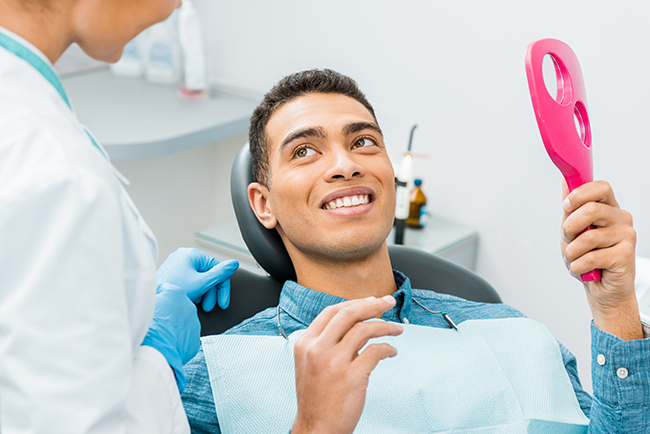
Regular oral exams are the simplest, most effective way to preserve a healthy mouth and to spot issues while they remain easy to manage. At the office of Skyline Smiles, our goal during each exam is to combine careful observation, appropriate imaging, and clear communication so you understand your oral health and the reasoning behind any recommendations. If you have questions about what happens during an exam or whether certain screenings or images are right for you, please contact us for more information.
A routine oral exam is a systematic evaluation of your teeth, gums, and surrounding tissues designed to catch problems early and document change over time. The clinician begins with a review of your medical and dental history and a short conversation about any symptoms such as sensitivity, soreness, or changes in bite. That intake informs a hands-on intraoral and extraoral inspection that looks for decay, wear, pocketing, and soft-tissue irregularities.
Exams also include an oral cancer screening of the mouth, throat, and neck and an assessment of jaw movement and bite relationships. When necessary, the clinician will recommend targeted imaging such as digital radiographs or photographs to look beneath the surface. Findings are explained clearly so you understand what was observed and what sensible next steps are recommended.
Before your first comprehensive exam, bring a list of current medications, recent medical changes, and any dental concerns you want addressed. If you have dental records or recent x-rays from another office, bring them or request they be transferred to your chart to provide historical context. Arrive a few minutes early to complete intake forms and to ensure the team has time to review your history.
Be ready to discuss habits that affect oral health, such as tobacco use, grinding, or dietary patterns, because these influence risk assessment and preventive recommendations. The clinical exam will be tailored to the information you share, so candid answers help the team identify issues that might be missed in a visual inspection alone. This preparation makes the visit more efficient and helps the clinician create a personalized care plan.
Regular oral exams detect dental and periodontal disease in early stages when treatment is simpler and more predictable. Many oral conditions progress silently; periodic professional evaluation reveals changes in tooth integrity, gum health, and soft tissues before symptoms become severe. Maintaining consistent records also allows clinicians to compare images and findings over time, improving diagnostic accuracy.
Oral health is linked to general health because signs of systemic conditions often appear in the mouth, including effects from medications, autoimmune disorders, and nutritional deficiencies. Detecting these signs early prompts appropriate referrals or conversations with your medical providers. Preventive exams therefore protect both your smile and broader wellbeing by encouraging timely intervention and coordinated care.
An oral cancer screening is a focused inspection of the lips, tongue, floor of mouth, cheeks, throat, and neck for persistent changes such as lumps, ulcers, or color variations. The clinician palpates tissues to detect masses or firm areas and visually inspects mucosal surfaces for sores or patches that do not heal. Screenings are performed as a standard part of comprehensive exams because early detection greatly improves outcomes.
If a suspicious area is found, the team documents its location and appearance, discusses next steps, and may recommend further testing or a referral to a specialist. Documentation and follow-up allow clinicians to monitor changes over time and determine whether biopsy or additional evaluation is warranted. Clear communication ensures you understand the reason for any recommendation and the expected timeline for follow-up.
Imaging is recommended based on clinical findings, symptoms, and risk factors rather than automatically at every visit. Bitewing radiographs are commonly used to check for interproximal decay and monitor bone levels, while periapical films target the root and surrounding bone of a specific tooth. A full-mouth series or panoramic image may be advised to establish a baseline or to assess broad anatomical concerns when indicated.
Digital systems minimize radiation exposure and provide immediate images that can be enlarged and enhanced for patient education and record-keeping. For complex planning, such as implant placement or assessment of anatomical structures, three-dimensional cone-beam scans may be suggested to improve precision. Your provider will explain the purpose of any recommended image so you understand what it shows and why it is helpful for your care.
The choice of radiograph depends on the clinical question that needs answering, the area of concern, and the patient’s risk profile. Bitewings are efficient for detecting decay between teeth and monitoring crestal bone, while periapicals are selected for detailed views of a single tooth and its root. Panoramic images provide an overview of the jaws and teeth and are useful for growth assessment, broad structural evaluation, or surgical planning.
CBCT scans are reserved for cases that require three-dimensional detail, such as implant planning or evaluation of complex anatomy. Clinicians prioritize targeted imaging that answers a specific diagnostic question and avoid unnecessary exposure by not over-imaging. Any recommended film or scan will be described in plain language so you know what information it will provide.
Oral exams are tailored to the patient’s stage of life and specific risks; children are evaluated for developmental milestones, eruption patterns, and early signs that might indicate future orthodontic needs. For adults, exams focus on detecting decay, periodontal disease, wear patterns from grinding, and soft-tissue changes related to lifestyle or medication. Older adults require attention to gum recession, prosthetic needs, root decay, and conditions that affect saliva and healing.
The frequency and scope of imaging and preventive measures are adjusted according to age and risk factors, with an emphasis on education and habit modification across all groups. Continuity of care helps clinicians track developmental or degenerative trends so interventions can be planned conservatively. This lifespan approach supports preventive dentistry and reduces the likelihood of emergency or complex treatment later on.
The hygienist performs a clinical assessment of periodontal health, measures pocket depths, and removes hardened plaque and tartar that brushing and flossing cannot eliminate. During the appointment they also evaluate tissue appearance, note areas of recession or inflammation, and document findings that help the dentist complete a comprehensive exam. Professional cleaning reduces inflammation and helps prevent progression of gum disease.
Hygienists provide practical education on brushing and interdental techniques, recommend appropriate home-care products, and suggest behavioral changes that protect oral health. Their observations contribute to the patient record and guide the dentist’s diagnostic decisions and treatment priorities. Working together, the hygienist and clinician create a plan that balances preventive care with any necessary restorative or periodontal follow-up.
Monitoring periodontal health begins with baseline measurements of pocket depths, attachment levels, and noted areas of bleeding or recession, all of which are recorded in the chart. Subsequent exams compare those measurements and clinical photos or radiographs to detect progression or stability. Regular professional cleanings and targeted interventions are scheduled according to the disease level and response to treatment.
When pockets, bone loss, or persistent inflammation are detected, clinicians may recommend enhanced hygiene protocols, more frequent maintenance visits, or referrals to periodontal specialists for additional care. Ongoing measurement and imaging allow the team to evaluate treatment effectiveness and adjust the plan as needed. Clear documentation and patient education are central to managing periodontal conditions successfully.
For most patients, routine oral exams and cleanings every six months provide an effective schedule for prevention and early detection, though frequency can be adjusted based on individual risk factors. Patients with high caries risk, active periodontal disease, or complex restorative needs may require more frequent monitoring and maintenance visits. The clinician will recommend a follow-up interval that reflects your risk profile and treatment goals.
You should schedule an earlier visit if you experience new pain, swelling, loose teeth, a sore that does not heal, sudden changes in bite, or other concerning symptoms. Early evaluation of acute changes helps prevent small problems from becoming more complex and supports timely coordination with medical providers when systemic issues may be involved. If you have questions about timing or specific screenings, contact the office of Skyline Smiles to discuss what schedule is right for you.
Whether you are due for a cleaning or finally ready to fix something that has bothered you for years, the first step is the same. Pick the office nearest you and we will find a time that works.
